
Male internal reproductive pathology model with anatomical accuracy shows urethral knots, benign prostatic hyperplasia, sharp warts, spermatogonia liu and other disease manifestations for medical education and training


Male internal reproductive pathology model with anatomical accuracy shows urethral knots, benign prostatic hyperplasia, sharp warts, spermatogonia liu and other disease manifestations for medical education and training
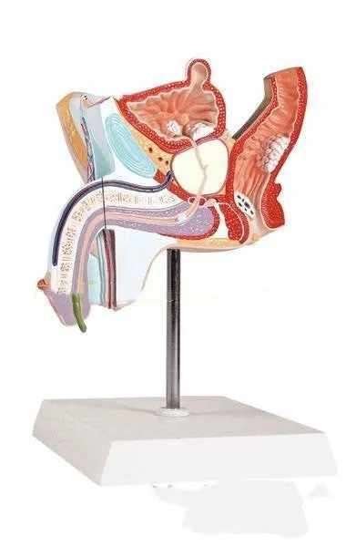
Male reproductive pathology anatomical model showing urethral knots, benign prostatic hyperplasia, and sharp warts

This anatomical model provides a detailed, three-dimensional representation of the male urinary and reproductive system, designed specifically to illustrate common pathological conditions. Constructed at a one-to-one ratio, it serves as an essential visual tool for medical students, educators, and healthcare professionals seeking to understand the anatomical presentation of diseases such as benign prostatic hyperplasia, urethral knots, and sharp warts. Its clear design allows for focused study of these conditions within their correct anatomical context, making it a practical resource for both classroom instruction and clinical training environments where visual aids enhance comprehension.
Features and Construction
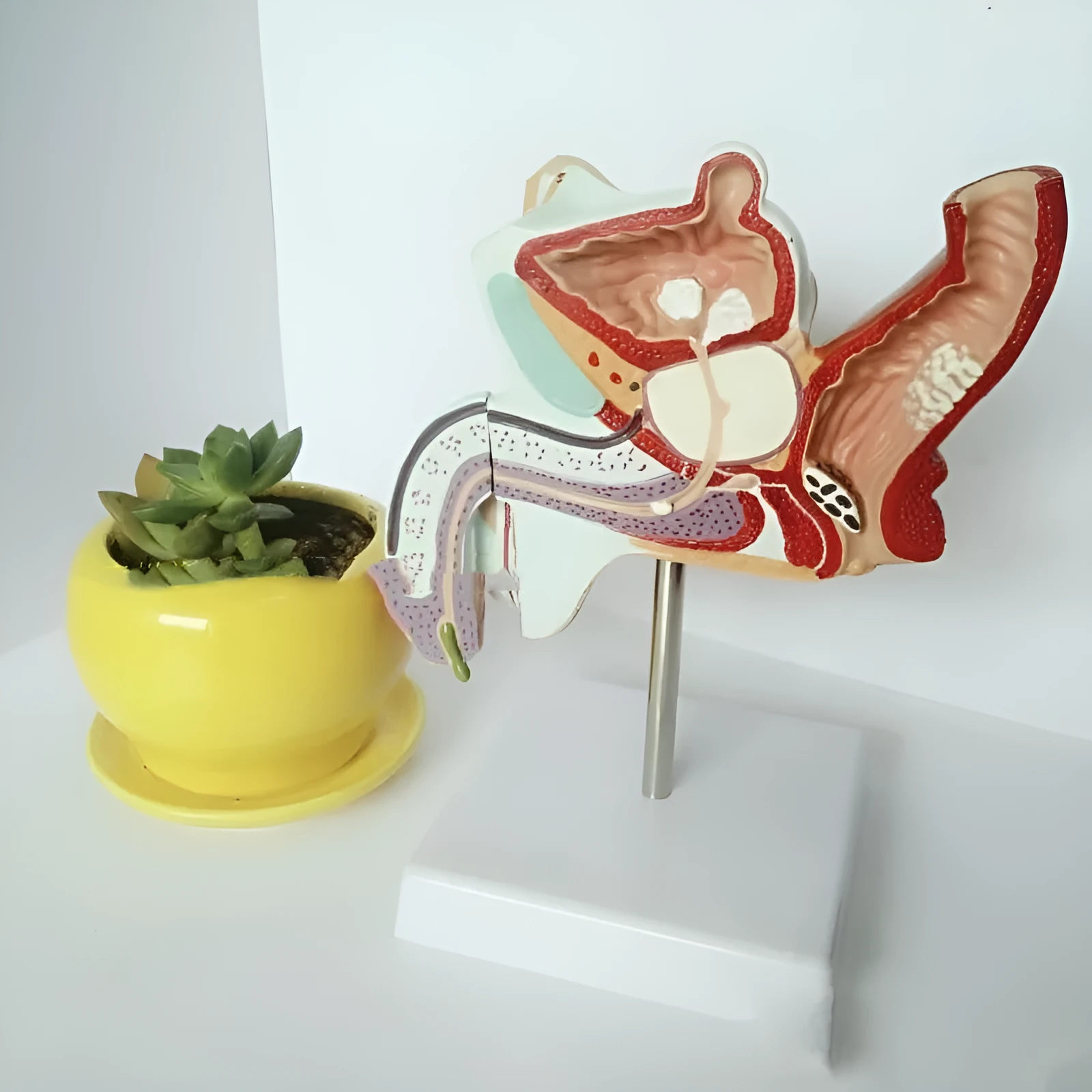
The model’s design centres on anatomical accuracy and educational utility. It is built to show multiple pathologies simultaneously, facilitating comparative study and a comprehensive overview of male reproductive system diseases.
Material and Build
Fabricated from plastic, the model offers a durable construction suitable for repeated handling in educational settings. The material allows for the precise moulding of anatomical details necessary to represent the pathological states, including the distinct visual characteristics of urethral knots, benign prostatic hyperplasia, and sharp warts. The use of plastic contributes to the model's longevity as a teaching aid.
Size and Practical Fit
With dimensions of 25.5cm in width, 18cm in height, and 15cm in depth, the model provides a sufficiently detailed scale for individual or small-group study. Its size makes it portable and easy to place on a desk, examination table, or demonstration shelf. The one-to-one ratio design ensures that the spatial relationships between anatomical structures and pathologies are accurately maintained for effective learning.
Uses and Placement
This model is intended for scenarios where a tangible, visual explanation of male reproductive pathologies is required. Its application spans formal education, professional training, and patient consultation contexts.
Event or Professional Use
In medical schools and nursing programmes, the model serves as a standard teaching aid for urology and reproductive health modules. Healthcare professionals can use it during training seminars or in clinical settings to explain conditions like benign prostatic hyperplasia to patients or junior staff. Its clear depiction of multiple diseases makes it a versatile tool for comparative pathology lessons and diagnostic training.
Everyday Home Use
While primarily designed for educational and professional environments, the model may also be used by individuals studying anatomy or pathology independently. Its detailed construction supports self-directed learning for students preparing for exams or professionals seeking a refresher on male reproductive system diseases.
Benefits and Buying Value

The model’s value lies in its specific focus on pathology and its practical application in knowledge transfer. It consolidates the visual study of several conditions into a single, durable resource.
Reuse and Low Maintenance
The plastic construction allows the model to be reused repeatedly across multiple teaching sessions or academic years. Basic care involves wiping the surface with a damp cloth to remove dust, requiring minimal maintenance. This durability supports its role as a long-term investment for institutions or educators.
Why Choose This Product
This model is selected for its direct representation of specific male reproductive pathologies—urethral knots, benign prostatic hyperplasia, sharp warts, and spermatogonia liu—within an accurate anatomical framework. Its one-to-one ratio and clear detailing make it a focused tool for anyone needing to visualise these conditions, distinguishing it from more general anatomical models. It meets the need for a specialised, factual teaching aid in a field where accurate visualisation aids understanding.